High-Content Imaging Systems
High-Content Imaging (HCI) systems, sometimes associated with High-Content Screening (HCS), also known as High-Content Analysis (HCA), integrate advanced microscopy with automated image acquisition for cell imaging and quantitative analysis to extract rich cellular information from complex biological samples. Unlike HCS, which applies these capabilities to large-scale screening workflows, HCI focuses on the precise imaging and measurement of multiple cellular parameters such as morphology, protein localization, and signaling events. These systems employ fluorescence and confocal microscopy to capture detailed, high-resolution images efficiently. They are highly adaptable and compatible with various sample types, including adherent and suspension cells, organoids, and 3D cultures. Integrated analysis software automates image processing and data quantification, reducing manual workload, improving reproducibility, and enabling deeper insights into cellular behavior.read more
Key Features of High-Content Imaging Systems
- The systems enable complete automation of plate and slide imaging, including vessel handling and scanning. Motorized stages, filter cubes, and objective turrets allow precise and reproducible positioning, minimizing variability across experiments.
- Equipped with high-performance optics and laser autofocus, they capture high-resolution images rapidly while reducing phototoxicity and photobleaching. The optimized illumination and focus mechanisms ensure consistent image quality across different sample types.
- Integrated onstage incubation systems allow experiments under physiological and non-physiological conditions. This enables long-term live-cell imaging and monitoring of dynamic cellular processes without compromising cell viability.
- HCI systems provide several imaging modes, including multichannel fluorescence, brightfield, color brightfield, and phase contrast. This versatility allows researchers to capture diverse cellular features in a single workflow.
- Powerful software allows fully customizable analysis and quantification across multiple image-based phenotypes. Users can automate image processing, extract multiparametric data, and apply AI-assisted tools for deeper insights.
- The systems feature modular designs that can be tailored to current research needs and upgraded as requirements evolve. Flexible configurations support a range of sample types, from 2D cultures to complex 3D models.
- High-content imaging systems can integrate with robotic plate handlers, liquid handling modules, and other automated workflows. This improves throughput, reduces manual labor, and ensures reproducible and reliable data acquisition.
Applications
- Cell biology and Drug discovery: The technology supports understanding how drugs interact with cellular components and regulate biological functions. Observing compound effects on pathways and cellular processes provides critical insights into mechanisms of action.
- Assay optimization: High-content imaging is instrumental in designing and refining cellular assays. By enabling detailed visualization and quantitative analysis of cellular features, it helps researchers create robust assays that accurately measure specific biological activities.
- Phenotype-based screening: The high-content imaging system is valuable for identifying compounds that induce specific cellular phenotypes. By analyzing morphology, protein distribution, and other phenotypic traits, it aids in discovering drugs with desired effects or novel targets for therapeutic development.
- Gene delivery assessment: The system is extensively applied in transfection experiments to evaluate the efficiency and effects of introducing genetic material into cells. By tracking and quantifying gene expression, it helps optimize transfection protocols, study gene function, and investigate molecular interactions within the cell.
- Drug compound evaluation: Initially developed to complement high-throughput screening, high-content imaging allows rapid testing of large compound libraries. It evaluates effects on cell morphology, protein expression, and subcellular localization across multiple parameters simultaneously, facilitating the identification of promising drug candidates.
- Toxicity and safety profiling: The high-content imaging instrument is widely used to evaluate the safety of drug candidates and environmental agents. It enables monitoring of cell viability, apoptosis, oxidative stress, and other toxicological endpoints, helping to identify compounds or conditions that may negatively affect cells.
- Cellular cytometry analysis: The technology aids in quantifying various cellular characteristics, including cell cycle progression, DNA content, nuclear morphology, and chromatin organization. It allows the detection of abnormal cell populations and the study of cellular heterogeneity.
- Cancer cell studies: High-content imagers are pivotal in cancer research, enabling the analysis of cell behavior, tumor progression, and treatment responses. They facilitate the study of proliferation, migration, invasion, apoptosis, and the identification of potential therapeutic targets.
- Dynamic live cell monitoring: The instrument supports real-time observation of living cells, capturing processes such as migration, cell-cell interactions, and signaling events. Time-lapse imaging allows researchers to track changes in cellular phenotypes over time, providing insights into cellular dynamics.
- Virology Research: High-content imaging is used to examine virus-host interactions, viral replication, and the effects of antiviral compounds. It enables visualization and quantification of infection dynamics, viral protein expression, and the impact of viruses on cellular phenotypes.
- Neuronal analysis: In neuroscience, high-content imaging allows the study of neuronal structure, synapse formation, and activity. It aids in evaluating neuronal networks, neurite extension, and dendritic spine dynamics, contributing to research on neurodevelopmental and neurodegenerative disorders.
- Stem cell characterization: It is critical for stem cell research, allowing detailed analysis of stem cell identity, differentiation, and tissue engineering processes. It supports monitoring of pluripotency markers, cell fate decisions, and optimization of culture conditions.
- Cell biology insights: High-content imaging advances cell biology by enabling the study of protein localization, signaling pathways, and cellular processes. It provides comprehensive information on cell structure and function, facilitating the understanding of biological mechanisms and disease.
- Personalized therapeutics: The technology can be applied to personalized medicine by assessing patient-specific cellular responses to treatments. Analysis of cellular behavior and drug responses can guide individualized therapy decisions and optimize treatment strategies.
Showing 1–12 of 20 results
Category
- Bioreactors+
- Parallel Bioreactors
- Single-Use Bioreactors
- Stirred-Tank Bioreactors
- Capillary Electrophoresis Instruments+
- Capillary Gel Electrophoresis (CGE)
- Capillary Isoelectric Focusing (CIEF)
- Capillary Zone Electrophoresis (CZE)
- Cell Counters and Analyzers+
- Automated Cell Counters
- Manual Cell Counters
- Cellular Imaging Systems+
- Fluorescence Imaging Systems
- High-Content Imaging Systems
- Live-Cell Imaging Systems
- Centrifuges+
- Benchtop Centrifuges
- Clinical Centrifuges
- Micro Centrifuges
- Refrigerated Centrifuges
- Ultracentrifuges
- Chromatography Instruments+
- Gas Chromatography
- Ion Chromatography
- Liquid Chromatography
- Flow Cytometer+
- Cell Analyzers
- Cell Isolation Systems
- Cell Sorters
- High-Throughput Flow Cytometers
- Imaging Flow Cytometry
- Personal Flow Cytometers
- Portable/Benchtop Flow Cytometers
- Spectral Flow Cytometers
- Laboratory Incubators+
- CO2 Incubators
- Dry Bath Incubators
- Hypoxia Incubators and Chambers
- Incubator Shakers
- Laboratory Ovens
- Portable Incubators
- Thermal Mixers
- Laboratory Microscopes+
- Atomic Force Microscopes (AFM)
- Automated Modular Microscopes
- Compound Microscopes
- Confocal Microscopes
- Digital Microscopes
- Fluorescence Microscopes
- Inverted Microscopes
- Live Cell Imaging Microscopes
- Metallurgical Microscopes
- Multiphoton Microscopes
- Polarizing Microscopes
- Raman Microscopes
- Scanning Electron Microscopes (SEM)
- Stereo Microscopes
- Super-Resolution Microscopes
- Transmission Electron Microscopes (TEM)
- Upright Microscopes (Neuroscience)
- Liquid Handlers+
- Automated Pipetting Systems
- Multi-Channel Liquid Handlers
- Robotic Liquid Handlers
- Variable Volume Pipetting Systems
- Mass Spectrometers+
- Ambient Ionization
- APCI MS
- ESI MS
- FT ICR MS
- ICP MS
- Orbi Trap MS
- Quadrupole MS
- Time-of-Flight MS
- Microarray Scanners+
- DNA Microarray Scanners
- Laser-Based Microarray Scanners
- Microplate Readers+
- Absorbance Microplate Readers
- Fluorescence Microplate Readers
- Luminescence Microplate Readers
- Multimode Microplate Readers
- Particle Counters and Analyzers+
- Airborne Particle Counters
- Laser Diffraction Analyzers
- Liquid Particle Counters
- Nanoparticle Analyzers
- Portable Particle Counters
- Remote Particle Counters
Brand
- ACCU-SCOPE
- Agilent
- Airmodus
- Alphasense
- Analytik Jena
- ARGO-HYTOS
- Arrayit
- Aurora Biomed Inc.
- Azure Biosystems
- Baker
- BD Biosciences
- Beckman Coulter
- Beijing Challen Biotechnology
- Binder
- Bio-Techne
- Biobase
- BioRad
- BMG Labtech
- Bruker
- Celestron
- Cellbox
- Centurion Scientific Ltd
- ChemoMetec
- CleanAir
- Cleatech
- Corning
- Countstar
- CS Instruments
- CUBIC
- Cytek Biosciences
- Cytena
- Cytiva
- Data Technologies
- Descase
- Electrolab Biotech
- Eppendorf
- Euromex
- Evident
- Fermex
- Fluke
- Formulatrix
- Getinge
- Gilson
- GPC Bio
- Haier Biomedical
- Hamilton Company
- Hettich
- Hitachi High-Tech
- Hudson Robotics Inc.
- Hydro
- INFORS HT
- Innopsys
- Jasco
- Jeiotech
- JEOL
- Kanomax
- Keyence
- KNAUER
- LAB-KITS
- Labomed
- Labwit
- Leco
- Leica
- Lighthouse
- Logos Biosystems
- LOSI
- Lumex Instruments
- Luminex
- Malvern Panalytical
- Meiji Techno
- Memmert
- Merck
- Mettler-Toledo International Inc.
- Miltenyi Biotec
- Molecular Devices
- Motic
- MP Filtri
- MSE (Medical and Scientific Equipment)
- NCI
- Nikon Instruments
- NuAire
- ORFLO Technologies
- Oxford Instruments
- PAMAS
- Particle Measuring Systems
- Particles Plus
- PBS Biotech
- PCE instruments
- PerkinElmer
- PharSol
- PHCbi
- Prior Scientific
- Promega
- Qiagen NV
- Revvity
- RION
- Sartorius
- SATAKE
- Sciex
- Sebia
- SETA
- Setra
- Shimadzu
- SIBATA
- Sigma Laborzentrifugen GmbH
- Solaris Biotech
- Solida Biotech
- Sony Biotechnology Inc.
- SPT Labtech Ltd
- Stratedigm Inc.
- Sysmex Corporation
- Tecan
- TES
- Thermo Fisher Scientific
- TSI
- Waters
- Western States Machine Company
- ZEISS
- ZETRON
-

Agilent BioTek Cytation 1
The Cytation 1 offers multimode detection with optional imaging, suited for entry screening and simple assays
-

Agilent BioTek Cytation 5
Cytation 5 combines widefield imaging (fluorescence, brightfield) with sensitive multimode plate detection in compact design
-

Agilent BioTek Cytation 7
Cytation 7 offers flexible imaging (fluorescence, brightfield, phase contrast) plus absorbance/fluorescence detection and live-cell control
-
Agilent BioTek Cytation C10
The Cytation C10 integrates spinning-disk confocal and widefield imaging with multimode microplate detection, live-cell incubation and Gen5 software c...
-

Evident scanR
Fully automated image acquisition and AI‑driven analysis for biological samples
-

Molecular Devices ImageXpress HCS.ai
ImageXpress HCS AI enables high-throughput imaging with integrated AI analysis for improved insights into complex cellular data.
-

Molecular Devices ImageXpress Micro
ImageXpress Micro Confocal supports advanced high-content screening with customizable confocal imaging and automation-ready operation.
-

Molecular Devices ImageXpress Micro 4
ImageXpress Micro 4 delivers advanced high-content imaging with automation features designed for comprehensive cellular research and high-throughput a...
-

Molecular Devices ImageXpress Nano
ImageXpress Nano delivers automated high-content screening with brightfield and widefield fluorescence imaging, suitable for multi-parametric cellular...
-
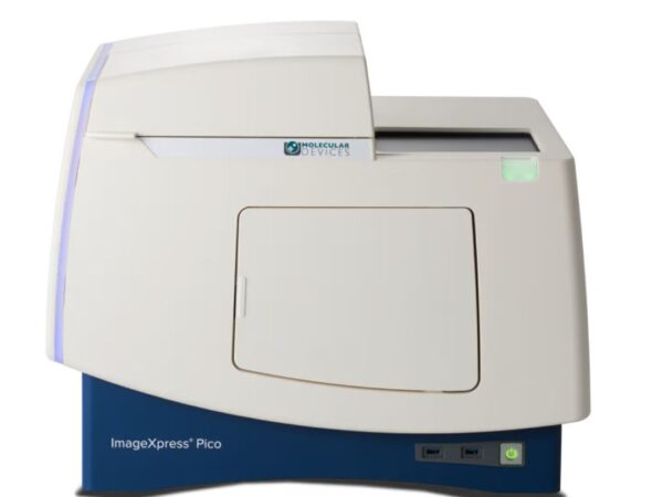
Molecular Devices ImageXpress Pico
ImageXpress Pico is a compact high-content imaging system with automation, robust analysis tools, and user-friendly interface ideal for research labs.
-

Nikon Instruments BioPipeline Live
A high-throughput live cell system with automated plate handling, customizable detectors, scheduler, and data handling server.
-

Nikon Instruments BioPipeline Plate
A high-content platform combining robotics, confocal modalities, scheduler and a large field-of-view for high-resolution screening.