Imaging Flow Cytometry
Imaging flow cytometry (IFC) is an advanced technique that merges flow cytometry’s ability to rapidly analyze large numbers of cells with microscopy’s ability to capture detailed images of each individual cell. By providing both quantitative data and spatial information, IFC enables researchers to study cell morphology, marker localization, and complex phenotypes at the single-cell level. It works by passing cells in a fluid stream through a focused beam of light, where high-speed cameras capture multiple images of each cell across different channels, such as brightfield, darkfield, and fluorescence. These images are then processed to extract hundreds of quantitative features, enabling detailed analysis of cell morphology, marker distribution, and phenotype classification at the single-cell level. Although traditional IFC has faced throughput limitations, recent technological innovations have dramatically increased imaging speed and processing capacity.read more
Essential Features
- Cells in suspension are hydrodynamically focused into a narrow stream and illuminated by an LED array combined with multiple laser lines. This setup enables imaging at rates of several thousand objects per second.
- A standard excitation laser is used, with the option to expand to multiple additional lasers at different wavelengths. Higher-power laser configurations are available for enhanced signal detection.
- The system can capture images across multiple channels, including several fluorescence channels along with brightfield and darkfield images. Dual detection systems with filters and spectral decomposition are used to increase the number of simultaneously acquired images.
- Images can be collected at different magnifications, typically corresponding to submicron pixel resolutions. This flexibility allows detailed visualization of both whole cells and fine subcellular structures.
- An optional extended depth-of-field setting preserves focus across greater cell thickness. This is useful for quantifying features throughout the entire volume of a cell.
- A high-gain configuration enhances the ability to detect weak fluorescence signals. It is particularly effective for very small or dim targets such as extracellular vesicles or viruses.
- User-friendly acquisition software enables instrument calibration, setup, and real-time monitoring of image data. It also supports gating strategies to selectively capture relevant events and reduce unnecessary data.
Applications of Image Analysis with Imaging Flow Cytometers
- Immunology and cell therapy: Imaging cytometry is used to study immune cell interactions, such as antigen-presenting cell binding, immune synapse formation, and CAR-T cell function. It also enables precise measurement of cell death, protein colocalization, and signaling events at the single-cell level.
- Cell biology and drug response: Researchers apply IFC to track processes like autophagy through LC3 clustering, nuclear localization of transcription factors, and cyclin-dependent cell cycle progression. It is also used to quantify apoptotic indices after drug treatments, providing high-content insights into cellular responses.
- Extracellular vesicles and small particle analysis: The technology allows visualization and quantification of extracellular vesicle uptake by cells. Its high sensitivity enables the detection of small or dim structures that may be missed by conventional cytometry.
- Aquatic microbiology and environmental monitoring: The image-based cytometer supports in situ analysis of plankton, bacteria, fungi, and other microorganisms in aquatic ecosystems. It helps detect ecological changes such as harmful algal blooms by monitoring different compartments simultaneously.
- Genotoxicity and toxicology: For assays like the micronucleus test, IFC offers a more accurate and high-throughput alternative to flow cytometry and microscopy. By imaging intact cells, it reduces false positives, enables counting of multiple micronuclei per cell, and supports automated AI-driven scoring for robust statistics.
- Nuclear translocation studies: Image-based flow cytometry enables precise quantification of nuclear translocation by simultaneously capturing fluorescence images of both the nucleus and the protein of interest within intact cells. This allows researchers to measure localization shifts objectively across thousands of cells, providing statistically robust insights into signaling pathways and transcription factor activation.
- Rare event imaging: Imaging flow cytometry is highly effective for rare event imaging, as it combines high-throughput acquisition with detailed imaging of each individual cell. This enables reliable detection and characterization of uncommon populations, such as circulating tumor cells or stem cell subsets, that may be overlooked using conventional methods.
Showing the single result
Category
- Bioreactors+
- Parallel Bioreactors
- Single-Use Bioreactors
- Stirred-Tank Bioreactors
- Capillary Electrophoresis Instruments+
- Capillary Gel Electrophoresis (CGE)
- Capillary Isoelectric Focusing (CIEF)
- Capillary Zone Electrophoresis (CZE)
- Cell Counters and Analyzers+
- Automated Cell Counters
- Manual Cell Counters
- Cellular Imaging Systems+
- Fluorescence Imaging Systems
- High-Content Imaging Systems
- Live-Cell Imaging Systems
- Centrifuges+
- Benchtop Centrifuges
- Clinical Centrifuges
- Micro Centrifuges
- Refrigerated Centrifuges
- Ultracentrifuges
- Chromatography Instruments+
- Gas Chromatography
- Ion Chromatography
- Liquid Chromatography
- Flow Cytometer+
- Cell Analyzers
- Cell Isolation Systems
- Cell Sorters
- High-Throughput Flow Cytometers
- Imaging Flow Cytometry
- Personal Flow Cytometers
- Portable/Benchtop Flow Cytometers
- Spectral Flow Cytometers
- Laboratory Incubators+
- CO2 Incubators
- Dry Bath Incubators
- Hypoxia Incubators and Chambers
- Incubator Shakers
- Laboratory Ovens
- Portable Incubators
- Thermal Mixers
- Laboratory Microscopes+
- Atomic Force Microscopes (AFM)
- Automated Modular Microscopes
- Compound Microscopes
- Confocal Microscopes
- Digital Microscopes
- Fluorescence Microscopes
- Inverted Microscopes
- Live Cell Imaging Microscopes
- Metallurgical Microscopes
- Multiphoton Microscopes
- Polarizing Microscopes
- Raman Microscopes
- Scanning Electron Microscopes (SEM)
- Stereo Microscopes
- Super-Resolution Microscopes
- Transmission Electron Microscopes (TEM)
- Upright Microscopes (Neuroscience)
- Liquid Handlers+
- Automated Pipetting Systems
- Multi-Channel Liquid Handlers
- Robotic Liquid Handlers
- Variable Volume Pipetting Systems
- Mass Spectrometers+
- Ambient Ionization
- APCI MS
- ESI MS
- FT ICR MS
- ICP MS
- Orbi Trap MS
- Quadrupole MS
- Time-of-Flight MS
- Microarray Scanners+
- DNA Microarray Scanners
- Laser-Based Microarray Scanners
- Microplate Readers+
- Absorbance Microplate Readers
- Fluorescence Microplate Readers
- Luminescence Microplate Readers
- Multimode Microplate Readers
- Particle Counters and Analyzers+
- Airborne Particle Counters
- Laser Diffraction Analyzers
- Liquid Particle Counters
- Nanoparticle Analyzers
- Portable Particle Counters
- Remote Particle Counters
Brand
- ACCU-SCOPE
- Agilent
- Airmodus
- Alphasense
- Analytik Jena
- ARGO-HYTOS
- Arrayit
- Aurora Biomed Inc.
- Azure Biosystems
- Baker
- BD Biosciences
- Beckman Coulter
- Beijing Challen Biotechnology
- Binder
- Bio-Techne
- Biobase
- BioRad
- BMG Labtech
- Bruker
- Celestron
- Cellbox
- Centurion Scientific Ltd
- ChemoMetec
- CleanAir
- Cleatech
- Corning
- Countstar
- CS Instruments
- CUBIC
- Cytek Biosciences
- Cytena
- Cytiva
- Data Technologies
- Descase
- Electrolab Biotech
- Eppendorf
- Euromex
- Evident
- Fermex
- Fluke
- Formulatrix
- Getinge
- Gilson
- GPC Bio
- Haier Biomedical
- Hamilton Company
- Hettich
- Hitachi High-Tech
- Hudson Robotics Inc.
- Hydro
- INFORS HT
- Innopsys
- Jasco
- Jeiotech
- JEOL
- Kanomax
- Keyence
- KNAUER
- LAB-KITS
- Labomed
- Labwit
- Leco
- Leica
- Lighthouse
- Logos Biosystems
- LOSI
- Lumex Instruments
- Luminex
- Malvern Panalytical
- Meiji Techno
- Memmert
- Merck
- Mettler-Toledo International Inc.
- Miltenyi Biotec
- Molecular Devices
- Motic
- MP Filtri
- MSE (Medical and Scientific Equipment)
- NCI
- Nikon Instruments
- NuAire
- ORFLO Technologies
- Oxford Instruments
- PAMAS
- Particle Measuring Systems
- Particles Plus
- PBS Biotech
- PCE instruments
- PerkinElmer
- PharSol
- PHCbi
- Prior Scientific
- Promega
- Qiagen NV
- Revvity
- RION
- Sartorius
- SATAKE
- Sciex
- Sebia
- SETA
- Setra
- Shimadzu
- SIBATA
- Sigma Laborzentrifugen GmbH
- Solaris Biotech
- Solida Biotech
- Sony Biotechnology Inc.
- SPT Labtech Ltd
- Stratedigm Inc.
- Sysmex Corporation
- Tecan
- TES
- Thermo Fisher Scientific
- TSI
- Waters
- Western States Machine Company
- ZEISS
- ZETRON
-
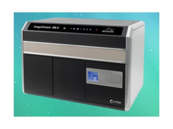
Cytek Biosciences Cytek® Amnis® ImageStream®X Mk II Imaging Flow Cytometer
ImageStream enables morphological and phenotypic analysis at single-cell resolution.