Confocal Microscopes
Confocal microscopes have become a central imaging platform for life science and biomedical research, offering the ability to visualize fine structural details in three dimensions with high contrast and clarity. Unlike conventional widefield instruments, a confocal microscope system uses focused point illumination and a pinhole to reject out-of-focus light, resulting in sharper images, improved optical sectioning, and more accurate quantitative measurements from thick or densely labeled samples. This makes confocal laser scanning microscopy particularly valuable when working with fluorescently labeled cells and tissues where precise localization of proteins, organelles, or signaling events is critical.read more
Researchers can acquire serial optical sections through their specimens and reconstruct detailed 3D image volumes, helping to reveal complex biological architecture such as neural networks, vasculature, and tissue microenvironments that are not easily resolved with standard fluorescence microscopes. Modern confocal microscope systems span a wide range of configurations, from basic confocal microscopy setups aimed at routine imaging through to advanced laser scanning and spinning disk platforms optimized for high-speed live-cell experiments, multiplexed imaging, and deep tissue investigations. This category brings together these confocal microscopes to help users compare capabilities and select systems that match their resolution, imaging depth, and workflow requirements.
Key Features
- High-resolution optical sectioning: Confocal microscopes capture optical sections from thick specimens without physical sectioning, achieving lateral resolution of approximately 0.2 μm and axial resolution around 0.5-0.6 μm.
- Superior out-of-focus light rejection: Point-like illumination and spatial filtering through pinholes eliminate out-of-focus light, resulting in superior image clarity compared to widefield microscopes.
- Three-dimensional reconstruction capability: Systems generate thin optical slices through specimens by rejecting light from above and below the focal plane, enabling 3D reconstruction of cellular and tissue architecture.
- Adjustable optical section thickness: Optical section depth is controlled by numerical aperture of objective lenses and pinhole size, allowing customization based on specimen requirements.
- Multi-color fluorescence imaging: Modern confocal microscope systems offer multi-color imaging capabilities and compatibility with both live and fixed samples.
- Laser scanning confocal microscopy (LSCM): Uses single pinhole scanning point-by-point across specimens, providing optimal resolution and flexibility for fixed tissue imaging.
- Spinning disk confocal systems: Employ hundreds of pinholes rotating at high speed for simultaneous imaging across the entire field with reduced photodamage for live-cell applications.
Applications of Confocal Microscopes
- Biomedical tissue imaging: Enables rapid examination of thick tissue specimens with fluorescence imaging to investigate three-dimensional distributions of cellular structures, including nerve networks and vascular patterns.
- Immunofluorescence histochemistry: Combines confocal microscopy with immunofluorescence techniques for detailed analysis of protein localization and cell-cell interactions without physical sectioning.
- Live-cell imaging: Basic confocal microscopy techniques monitor dynamic cellular processes in real time with minimal photobleaching and photodamage during extended observation periods.
- Intracellular ion flux measurements: Quantitative measurements of calcium signaling and other intracellular ion fluxes using rapid line-scanning confocal configurations.
- Materials science characterization: Non-destructive characterization of complex three-dimensional structures in materials research applications.
- Developmental biology studies: Supports investigations requiring high-contrast visualization of fluorescently labeled specimens in embryonic and developmental processes.
- Neuroscience research: Enables detailed mapping of neural architecture and connectivity in brain tissue and nervous system specimens.
- Pharmaceutical research: Facilitates drug discovery workflows requiring precise visualization of cellular responses and molecular interactions in both fixed and living samples.
Showing all 8 results
Category
- Bioreactors+
- Parallel Bioreactors
- Single-Use Bioreactors
- Stirred-Tank Bioreactors
- Capillary Electrophoresis Instruments+
- Capillary Gel Electrophoresis (CGE)
- Capillary Isoelectric Focusing (CIEF)
- Capillary Zone Electrophoresis (CZE)
- Cell Counters and Analyzers+
- Automated Cell Counters
- Manual Cell Counters
- Cellular Imaging Systems+
- Fluorescence Imaging Systems
- High-Content Imaging Systems
- Live-Cell Imaging Systems
- Centrifuges+
- Benchtop Centrifuges
- Clinical Centrifuges
- Micro Centrifuges
- Refrigerated Centrifuges
- Ultracentrifuges
- Chromatography Instruments+
- Gas Chromatography
- Ion Chromatography
- Liquid Chromatography
- Flow Cytometer+
- Cell Analyzers
- Cell Isolation Systems
- Cell Sorters
- High-Throughput Flow Cytometers
- Imaging Flow Cytometry
- Personal Flow Cytometers
- Portable/Benchtop Flow Cytometers
- Spectral Flow Cytometers
- Laboratory Incubators+
- CO2 Incubators
- Dry Bath Incubators
- Hypoxia Incubators and Chambers
- Incubator Shakers
- Laboratory Ovens
- Portable Incubators
- Thermal Mixers
- Laboratory Microscopes+
- Atomic Force Microscopes (AFM)
- Automated Modular Microscopes
- Compound Microscopes
- Confocal Microscopes
- Digital Microscopes
- Fluorescence Microscopes
- Inverted Microscopes
- Live Cell Imaging Microscopes
- Metallurgical Microscopes
- Multiphoton Microscopes
- Polarizing Microscopes
- Raman Microscopes
- Scanning Electron Microscopes (SEM)
- Stereo Microscopes
- Super-Resolution Microscopes
- Transmission Electron Microscopes (TEM)
- Upright Microscopes (Neuroscience)
- Liquid Handlers+
- Automated Pipetting Systems
- Multi-Channel Liquid Handlers
- Robotic Liquid Handlers
- Variable Volume Pipetting Systems
- Mass Spectrometers+
- Ambient Ionization
- APCI MS
- ESI MS
- FT ICR MS
- ICP MS
- Orbi Trap MS
- Quadrupole MS
- Time-of-Flight MS
- Microarray Scanners+
- DNA Microarray Scanners
- Laser-Based Microarray Scanners
- Microplate Readers+
- Absorbance Microplate Readers
- Fluorescence Microplate Readers
- Luminescence Microplate Readers
- Multimode Microplate Readers
- Particle Counters and Analyzers+
- Airborne Particle Counters
- Laser Diffraction Analyzers
- Liquid Particle Counters
- Nanoparticle Analyzers
- Portable Particle Counters
- Remote Particle Counters
Brand
- ACCU-SCOPE
- Agilent
- Airmodus
- Alphasense
- Analytik Jena
- ARGO-HYTOS
- Arrayit
- Aurora Biomed Inc.
- Azure Biosystems
- Baker
- BD Biosciences
- Beckman Coulter
- Beijing Challen Biotechnology
- Binder
- Bio-Techne
- Biobase
- BioRad
- BMG Labtech
- Bruker
- Celestron
- Cellbox
- Centurion Scientific Ltd
- ChemoMetec
- CleanAir
- Cleatech
- Corning
- Countstar
- CS Instruments
- CUBIC
- Cytek Biosciences
- Cytena
- Cytiva
- Data Technologies
- Descase
- Electrolab Biotech
- Eppendorf
- Euromex
- Evident
- Fermex
- Fluke
- Formulatrix
- Getinge
- Gilson
- GPC Bio
- Haier Biomedical
- Hamilton Company
- Hettich
- Hitachi High-Tech
- Hudson Robotics Inc.
- Hydro
- INFORS HT
- Innopsys
- Jasco
- Jeiotech
- JEOL
- Kanomax
- Keyence
- KNAUER
- LAB-KITS
- Labomed
- Labwit
- Leco
- Leica
- Lighthouse
- Logos Biosystems
- LOSI
- Lumex Instruments
- Luminex
- Malvern Panalytical
- Meiji Techno
- Memmert
- Merck
- Mettler-Toledo International Inc.
- Miltenyi Biotec
- Molecular Devices
- Motic
- MP Filtri
- MSE (Medical and Scientific Equipment)
- NCI
- Nikon Instruments
- NuAire
- ORFLO Technologies
- Oxford Instruments
- PAMAS
- Particle Measuring Systems
- Particles Plus
- PBS Biotech
- PCE instruments
- PerkinElmer
- PharSol
- PHCbi
- Prior Scientific
- Promega
- Qiagen NV
- Revvity
- RION
- Sartorius
- SATAKE
- Sciex
- Sebia
- SETA
- Setra
- Shimadzu
- SIBATA
- Sigma Laborzentrifugen GmbH
- Solaris Biotech
- Solida Biotech
- Sony Biotechnology Inc.
- SPT Labtech Ltd
- Stratedigm Inc.
- Sysmex Corporation
- Tecan
- TES
- Thermo Fisher Scientific
- TSI
- Waters
- Western States Machine Company
- ZEISS
- ZETRON
-
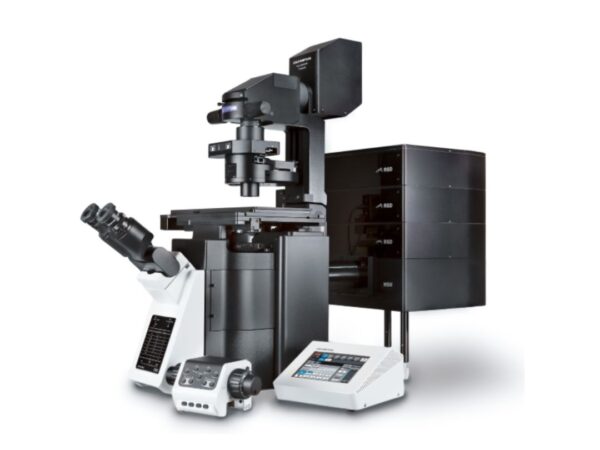
Evident FV4000
The FV4000 confocal system delivers fast acquisition, high resolution, and sensitivity for precise imaging.
-
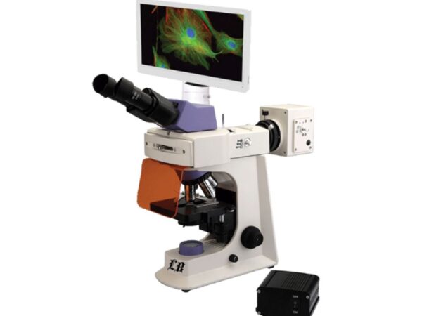
Labomed LB-1750
Offers reliable imaging performance in various lab environments.
-
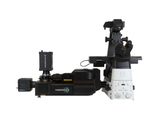
Nikon Instruments CrestOptics X-Light Series
Crest X-Light series offers flexible spinning disk confocal imaging solutions, ideal for high-throughput and multi-well plate applications.
-
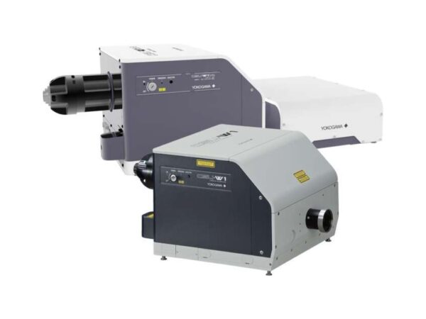
Nikon Instruments CSU
CSU series microscopes enable rapid, low-phototoxicity confocal imaging for real-time observation of cellular dynamics.
-
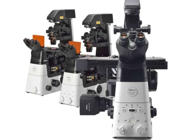
Nikon Instruments ECLIPSE Ti2-A
The Ti2-E platform offers automated imaging with superior stability, ideal for time-lapse and high-throughput workflows
-

Nikon Instruments ECLIPSE Ti2-E/Ti2-E/B*1
The Ti2-E platform offers automated imaging with superior stability, ideal for time-lapse and high-throughput workflows
-

Nikon Instruments ECLIPSE Ti2-U
The Ti2-E platform offers automated imaging with superior stability, ideal for time-lapse and high-throughput workflows
-

Nikon Instruments ECLIPSE Ti2-U IVF
The Ti2-U IVF system provides optimal optics and ergonomic features for IVF laboratories and clinics