Multiphoton Microscopes: Precision Imaging for Biopharma & Drug Discovery
In the rapidly evolving landscape of biopharmaceutical research, multiphoton microscopy has established itself as a critical imaging technique for visualizing complex biological mechanisms within deep tissue environments. Unlike traditional confocal microscopy, which makes use of single-photon excitation and often struggles with significant light scattering in thick specimens, multiphoton microscopes leverage two-photon excitation to penetrate hundreds of micrometers into dense biological matrices. This advanced form of fluorescence microscopy relies on the nonlinear optical process, where a fluorophore undergoes the simultaneous absorption of two photons to reach an excited state. This specific interaction is often described as a resonance phenomenon, occurring only when the combined energy of the photons matches the transition gap of the molecule. Because this absorption of two photons depends on the square of the excitation intensity, the fluorescence is confined strictly to the focal volume, inherently creating optical sectioning without the need for a pinhole to reject out-of-focus light.read more
For researchers in drug discovery, this capability is transformative. It allows for the high-resolution image capture of living cells and tissues within their native physiological context, a requirement for accurate pharmacokinetic and toxicological assessments. The necessary high photon density is achieved at the focal point by means of a pulsed laser system, typically a mode-locked laser emitting femtosecond pulses, maintaining a low average power simultaneously. This approach minimizes photobleaching and photodamage outside the focal plane, thereby preserving the viability of living tissue and live animals during longitudinal studies. Consequently, in vivo two-photon imaging has become the gold standard for intravital studies and the analysis of 3D cell culture models, such as spheroids and organoids, where standard optical methods fail to provide adequate penetration depth.
Category Highlights: Robust Performance for Preclinical Screening
The integration of multiphoton microscopes into pharmaceutical workflows addresses the limitations of conventional fluorescence microscopes by providing the stability and resolution necessary for preclinical screening. These systems are engineered to optimize the detection of scattered photons, ensuring that valuable signal from deep within the specimen is not lost.
- Deep Tissue Penetration: These microscopes minimize scattering and absorption via an excitation wavelength in the near-infrared light spectrum, which is a significantly longer wavelength than visible light. This allows for two-photon fluorescence imaging at depths exceeding 500 micrometers, enabling the visualization of the core of large tumor spheroids or intact organs.
- Minimized Phototoxicity: The multiphoton excitation process is spatially confined, meaning that photodamage and photobleaching are virtually non-existent outside the focal volume. This is critical for using two-photon microscopy in long-term live cell experiments, ensuring that the two-photon excited sample remains physiologically stable.
- Versatile Fluorophore Excitation: A single tunable laser source can often excite a fluorophore or multiple fluorescent protein variants simultaneously due to broad two-photon absorption spectra. This simplifies the optical setup for multi-color experiments tracking a drug and its target.
- Label-Free Modalities: Beyond fluorescence imaging, these systems support nonlinear contrast mechanisms such as fluorescence and second harmonic generation (SHG). This allows researchers to image collagen structures and fibrosis progression in living tissue without exogenous dyes.
- Enhanced Signal Collection: Since two-photon excitation microscopy does not require a confocal pinhole, the detectors can be placed as close to the objective as possible to capture all fluorescent emission, including scattered photons. This design maximizes sensitivity in turbid samples common in biopharma research.
Applications: Advancing Pharmacokinetics and Toxicology
The unique capabilities of two-photon microscopy make it indispensable for validating drug mechanisms and safety profiles. The technique bridges the gap between in vitro assays and clinical outcomes by providing high-fidelity data from in vivo and complex 3D models.
- Intravital Microscopy (IVM): Multiphoton laser scanning microscopy is extensively used for intravital imaging to monitor drug transport across biological barriers in live animals. Researchers can track the diffusion of therapeutic agents across the blood-brain barrier or into tumor vasculature in real-time, providing direct pharmacokinetic data.
- Neuroscience and Neurotoxicity: In the field of neurobiology, two-photon excitation is used to visualize structural plasticity, such as changes in dendritic spines, and functional activity in the neuron. This is vital for assessing the neuroprotective effects of new compounds or identifying potential neurotoxic side effects in the living brain.
- High-Content 3D Screening: As the industry shifts towards more predictive models, multiphoton imaging enables the detailed analysis of patient-derived organoids. It allows for the scan of entire 3D structures to evaluate drug efficacy and toxicity in a personalized medicine context.
- Metabolic Profiling: The metabolic state of cancer cells can be mapped via endogenous fluorophore signals like NADH by using fluorescence lifetime imaging (FLIM), in conjunction with multiphoton excitation. This lifetime imaging microscopy technique provides a label-free readout of cellular energetic shifts in response to treatment.
- Dermal Drug Delivery: The penetration depth afforded by using two-photon strategies is ideal for studying transdermal drug delivery. Researchers can visualize the distribution of topical formulations through the stratum corneum and into the deeper dermis.
Showing the single result
Category
- Bioreactors+
- Parallel Bioreactors
- Single-Use Bioreactors
- Stirred-Tank Bioreactors
- Capillary Electrophoresis Instruments+
- Capillary Gel Electrophoresis (CGE)
- Capillary Isoelectric Focusing (CIEF)
- Capillary Zone Electrophoresis (CZE)
- Cell Counters and Analyzers+
- Automated Cell Counters
- Manual Cell Counters
- Cellular Imaging Systems+
- Fluorescence Imaging Systems
- High-Content Imaging Systems
- Live-Cell Imaging Systems
- Centrifuges+
- Benchtop Centrifuges
- Clinical Centrifuges
- Micro Centrifuges
- Refrigerated Centrifuges
- Ultracentrifuges
- Chromatography Instruments+
- Gas Chromatography
- Ion Chromatography
- Liquid Chromatography
- Flow Cytometer+
- Cell Analyzers
- Cell Isolation Systems
- Cell Sorters
- High-Throughput Flow Cytometers
- Imaging Flow Cytometry
- Personal Flow Cytometers
- Portable/Benchtop Flow Cytometers
- Spectral Flow Cytometers
- Laboratory Incubators+
- CO2 Incubators
- Dry Bath Incubators
- Hypoxia Incubators and Chambers
- Incubator Shakers
- Laboratory Ovens
- Portable Incubators
- Thermal Mixers
- Laboratory Microscopes+
- Atomic Force Microscopes (AFM)
- Automated Modular Microscopes
- Compound Microscopes
- Confocal Microscopes
- Digital Microscopes
- Fluorescence Microscopes
- Inverted Microscopes
- Live Cell Imaging Microscopes
- Metallurgical Microscopes
- Multiphoton Microscopes
- Polarizing Microscopes
- Raman Microscopes
- Scanning Electron Microscopes (SEM)
- Stereo Microscopes
- Super-Resolution Microscopes
- Transmission Electron Microscopes (TEM)
- Upright Microscopes (Neuroscience)
- Liquid Handlers+
- Automated Pipetting Systems
- Multi-Channel Liquid Handlers
- Robotic Liquid Handlers
- Variable Volume Pipetting Systems
- Mass Spectrometers+
- Ambient Ionization
- APCI MS
- ESI MS
- FT ICR MS
- ICP MS
- Orbi Trap MS
- Quadrupole MS
- Time-of-Flight MS
- Microarray Scanners+
- DNA Microarray Scanners
- Laser-Based Microarray Scanners
- Microplate Readers+
- Absorbance Microplate Readers
- Fluorescence Microplate Readers
- Luminescence Microplate Readers
- Multimode Microplate Readers
- Particle Counters and Analyzers+
- Airborne Particle Counters
- Laser Diffraction Analyzers
- Liquid Particle Counters
- Nanoparticle Analyzers
- Portable Particle Counters
- Remote Particle Counters
Brand
- ACCU-SCOPE
- Agilent
- Airmodus
- Alphasense
- Analytik Jena
- ARGO-HYTOS
- Arrayit
- Aurora Biomed Inc.
- Azure Biosystems
- Baker
- BD Biosciences
- Beckman Coulter
- Beijing Challen Biotechnology
- Binder
- Bio-Techne
- Biobase
- BioRad
- BMG Labtech
- Bruker
- Celestron
- Cellbox
- Centurion Scientific Ltd
- ChemoMetec
- CleanAir
- Cleatech
- Corning
- Countstar
- CS Instruments
- CUBIC
- Cytek Biosciences
- Cytena
- Cytiva
- Data Technologies
- Descase
- Electrolab Biotech
- Eppendorf
- Euromex
- Evident
- Fermex
- Fluke
- Formulatrix
- Getinge
- Gilson
- GPC Bio
- Haier Biomedical
- Hamilton Company
- Hettich
- Hitachi High-Tech
- Hudson Robotics Inc.
- Hydro
- INFORS HT
- Innopsys
- Jasco
- Jeiotech
- JEOL
- Kanomax
- Keyence
- KNAUER
- LAB-KITS
- Labomed
- Labwit
- Leco
- Leica
- Lighthouse
- Logos Biosystems
- LOSI
- Lumex Instruments
- Luminex
- Malvern Panalytical
- Meiji Techno
- Memmert
- Merck
- Mettler-Toledo International Inc.
- Miltenyi Biotec
- Molecular Devices
- Motic
- MP Filtri
- MSE (Medical and Scientific Equipment)
- NCI
- Nikon Instruments
- NuAire
- ORFLO Technologies
- Oxford Instruments
- PAMAS
- Particle Measuring Systems
- Particles Plus
- PBS Biotech
- PCE instruments
- PerkinElmer
- PharSol
- PHCbi
- Prior Scientific
- Promega
- Qiagen NV
- Revvity
- RION
- Sartorius
- SATAKE
- Sciex
- Sebia
- SETA
- Setra
- Shimadzu
- SIBATA
- Sigma Laborzentrifugen GmbH
- Solaris Biotech
- Solida Biotech
- Sony Biotechnology Inc.
- SPT Labtech Ltd
- Stratedigm Inc.
- Sysmex Corporation
- Tecan
- TES
- Thermo Fisher Scientific
- TSI
- Waters
- Western States Machine Company
- ZEISS
- ZETRON
-
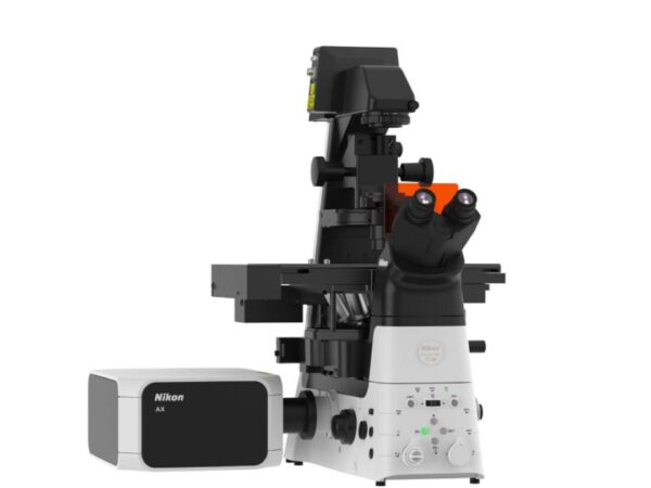
Nikon Instruments AX R with NSPARC
The AX with NSPARC delivers fast, high-resolution imaging with advanced photon detection for live-cell applications.