Super-Resolution Microscopes
Super-resolution microscopy has revolutionized the field of cellular and molecular imaging by breaking through the classical diffraction limit of conventional light microscopy. While traditional optical microscopes are constrained to resolutions of approximately 200-250 nm laterally and 500-700 nm axially, super-resolution technology now enables researchers to visualize biological structures at the nanometer scale, with some techniques achieving resolutions down to 5-20 nm. Recognized with the 2014 Nobel Prize in Chemistry, super-resolution microscopy encompasses several advanced techniques including Stimulated Emission Depletion (STED), Structured Illumination Microscopy (SIM), and single molecule localization microscopy methods such as Stochastic Optical Reconstruction Microscopy (STORM) and Photo-Activated Localization Microscopy (PALM). These powerful imaging modalities have transformed life sciences research by providing unprecedented insights into protein complexes, intracellular dynamics, and biomolecular structures that were previously impossible to observe. Super-resolution microscopy technique are now essential tools for researchers in molecular biology, cell biology, neuroscience, and drug discovery who require detailed visualization of cellular components and processes.read more
Key Features
Super-resolution microscopy systems offer distinct capabilities that enable researchers to overcome the limitations of conventional fluorescence microscopy:
- Enhanced spatial resolution: Super-resolution technology delivers lateral resolution limit ranging from 20-140 nm depending on the technique, compared to 200-250 nm for conventional microscopes, enabling visualization of previously unresolvable cellular structures.
- Multiple imaging modalities: Systems incorporate various super-resolution approaches including STED for confocal-based imaging, SIM for wide-field applications, and PALM/STORM for single-molecule localization microscopy.
- Three-dimensional imaging capabilities: Advanced super-resolution systems provide high-resolution 3D imaging technique with axial optical resolutions of 30-50 nm, allowing detailed reconstruction of cellular architectures.
- Live-cell compatibility: Techniques like SIM and certain STED configurations enable dynamic imaging of living cells, capturing real-time biological processes at the nanoscale.
- Multicolor super-resolution imaging: Modern systems support simultaneous imaging of multiple fluorescent labels, facilitating colocalization studies and protein interaction analyses at nanometer-scale resolution.
- Optimized detector specifications: High-sensitivity cameras with excellent signal-to-noise ratios are integrated to capture single-molecule emissions and low-intensity fluorescence signals critical for super high resolution microscopy.
- Laser stability and control: Precision laser systems with stable output and precise power modulation ensure consistent multicolor imaging performance during extended acquisition sessions.
- Flexible sample compatibility: Different super-resolution imaging modalities accommodate various sample types, from fixed cells to thick tissue sections and 3D biological specimens.
Applications of Super-Resolution Microscopes
Super-resolution microscopy has become an indispensable tool across multiple disciplines in life sciences, enabling researchers to investigate biological phenomena at unprecedented detail:
- Molecular and cellular biology: Researchers utilize super-resolution microscopy resolution capabilities to study protein localization, membrane organization, organelle structure, live cell imaging, and cytoskeletal dynamics with nanometer precision.
- Neuroscience research: Super resolution technology enables visualization of synaptic structures, neuronal connections, and neurotransmitter receptor distributions that are critical for understanding brain function and neurological disorders.
- Drug discovery and development: Pharmaceutical researchers employ super high resolution microscopy to examine drug-target interactions, cellular responses to therapeutic compounds, and disease-related structural changes at the molecular level.
- Chromatin and gene expression studies: Super-resolution techniques like PALM and STED reveal chromatin organization patterns, transcription factor binding sites, and nuclear architecture involved in gene regulation.
- Bacterial cell biology: Single-molecule imaging super-resolution microscopy provides insights into bacterial cytoskeleton structure, nucleoid organization, and dynamic processes of transcription and translation in prokaryotic systems.
- Protein complex analysis: Researchers apply super resolution electron microscopy and fluorescence-based approaches to characterize protein assemblies, molecular interactions, and biomolecular structures in vitro and in situ.
- Membrane biology and trafficking: Super-resolution imaging reveals membrane protein clustering, vesicle dynamics, endocytosis mechanisms, and lipid domain organization in cellular membranes.
- Tissue imaging and pathology: Advanced super-resolution systems enable detailed examination of tissue architecture, cellular interactions in three-dimensional environments, and pathological changes in disease states.
Showing all 2 results
Category
- Bioreactors+
- Parallel Bioreactors
- Single-Use Bioreactors
- Stirred-Tank Bioreactors
- Capillary Electrophoresis Instruments+
- Capillary Gel Electrophoresis (CGE)
- Capillary Isoelectric Focusing (CIEF)
- Capillary Zone Electrophoresis (CZE)
- Cell Counters and Analyzers+
- Automated Cell Counters
- Manual Cell Counters
- Cellular Imaging Systems+
- Fluorescence Imaging Systems
- High-Content Imaging Systems
- Live-Cell Imaging Systems
- Centrifuges+
- Benchtop Centrifuges
- Clinical Centrifuges
- Micro Centrifuges
- Refrigerated Centrifuges
- Ultracentrifuges
- Chromatography Instruments+
- Gas Chromatography
- Ion Chromatography
- Liquid Chromatography
- Flow Cytometer+
- Cell Analyzers
- Cell Isolation Systems
- Cell Sorters
- High-Throughput Flow Cytometers
- Imaging Flow Cytometry
- Personal Flow Cytometers
- Portable/Benchtop Flow Cytometers
- Spectral Flow Cytometers
- Laboratory Incubators+
- CO2 Incubators
- Dry Bath Incubators
- Hypoxia Incubators and Chambers
- Incubator Shakers
- Laboratory Ovens
- Portable Incubators
- Thermal Mixers
- Laboratory Microscopes+
- Atomic Force Microscopes (AFM)
- Automated Modular Microscopes
- Compound Microscopes
- Confocal Microscopes
- Digital Microscopes
- Fluorescence Microscopes
- Inverted Microscopes
- Live Cell Imaging Microscopes
- Metallurgical Microscopes
- Multiphoton Microscopes
- Polarizing Microscopes
- Raman Microscopes
- Scanning Electron Microscopes (SEM)
- Stereo Microscopes
- Super-Resolution Microscopes
- Transmission Electron Microscopes (TEM)
- Upright Microscopes (Neuroscience)
- Liquid Handlers+
- Automated Pipetting Systems
- Multi-Channel Liquid Handlers
- Robotic Liquid Handlers
- Variable Volume Pipetting Systems
- Mass Spectrometers+
- Ambient Ionization
- APCI MS
- ESI MS
- FT ICR MS
- ICP MS
- Orbi Trap MS
- Quadrupole MS
- Time-of-Flight MS
- Microarray Scanners+
- DNA Microarray Scanners
- Laser-Based Microarray Scanners
- Microplate Readers+
- Absorbance Microplate Readers
- Fluorescence Microplate Readers
- Luminescence Microplate Readers
- Multimode Microplate Readers
- Particle Counters and Analyzers+
- Airborne Particle Counters
- Laser Diffraction Analyzers
- Liquid Particle Counters
- Nanoparticle Analyzers
- Portable Particle Counters
- Remote Particle Counters
Brand
- ACCU-SCOPE
- Agilent
- Airmodus
- Alphasense
- Analytik Jena
- ARGO-HYTOS
- Arrayit
- Aurora Biomed Inc.
- Azure Biosystems
- Baker
- BD Biosciences
- Beckman Coulter
- Beijing Challen Biotechnology
- Binder
- Bio-Techne
- Biobase
- BioRad
- BMG Labtech
- Bruker
- Celestron
- Cellbox
- Centurion Scientific Ltd
- ChemoMetec
- CleanAir
- Cleatech
- Corning
- Countstar
- CS Instruments
- CUBIC
- Cytek Biosciences
- Cytena
- Cytiva
- Data Technologies
- Descase
- Electrolab Biotech
- Eppendorf
- Euromex
- Evident
- Fermex
- Fluke
- Formulatrix
- Getinge
- Gilson
- GPC Bio
- Haier Biomedical
- Hamilton Company
- Hettich
- Hitachi High-Tech
- Hudson Robotics Inc.
- Hydro
- INFORS HT
- Innopsys
- Jasco
- Jeiotech
- JEOL
- Kanomax
- Keyence
- KNAUER
- LAB-KITS
- Labomed
- Labwit
- Leco
- Leica
- Lighthouse
- Logos Biosystems
- LOSI
- Lumex Instruments
- Luminex
- Malvern Panalytical
- Meiji Techno
- Memmert
- Merck
- Mettler-Toledo International Inc.
- Miltenyi Biotec
- Molecular Devices
- Motic
- MP Filtri
- MSE (Medical and Scientific Equipment)
- NCI
- Nikon Instruments
- NuAire
- ORFLO Technologies
- Oxford Instruments
- PAMAS
- Particle Measuring Systems
- Particles Plus
- PBS Biotech
- PCE instruments
- PerkinElmer
- PharSol
- PHCbi
- Prior Scientific
- Promega
- Qiagen NV
- Revvity
- RION
- Sartorius
- SATAKE
- Sciex
- Sebia
- SETA
- Setra
- Shimadzu
- SIBATA
- Sigma Laborzentrifugen GmbH
- Solaris Biotech
- Solida Biotech
- Sony Biotechnology Inc.
- SPT Labtech Ltd
- Stratedigm Inc.
- Sysmex Corporation
- Tecan
- TES
- Thermo Fisher Scientific
- TSI
- Waters
- Western States Machine Company
- ZEISS
- ZETRON
-
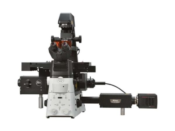
Nikon Instruments N-STORM
N-STORM enables precise localization microscopy for detailed molecular studies at the nanoscale level.
-
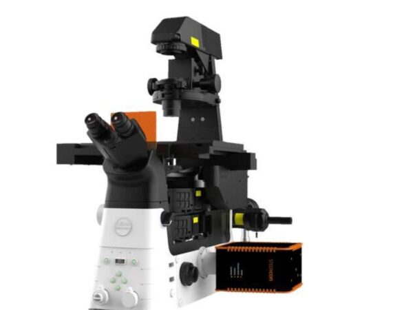
Nikon Instruments STEDYCON
STEDYCON combines STED microscopy precision with a compact design for easy integration with existing systems.