High-Fidelity Upright Microscope Systems for Clinical Sample Preparation, Fluorescence, and Light Microscopy in Life Sciences
The upright microscope remains the requisite optical platform for examining fixed biological specimens, acute tissue sections, and histological slides with high-fidelity resolution. Unlike inverted configurations, where the beam path converges from below, the upright microscope positions the lens above the specimen, facilitating superior immersion capabilities and optimizing the refractive index match for aqueous media.read more
This architecture is particularly crucial in life science disciplines requiring high numerical aperture (NA) visualization of opaque and semi-opaque samples, such as those typically encountered in clinical pathology and neuroanatomical mapping. Modern systems integrate motorized Z-axis control and infinity-corrected optics, ensuring that photon efficiency is maximized during fluorescence and brightfield microscopy. For researchers prioritizing sample preparation integrity, particularly when utilizing water-dipping optics for deep tissue imaging, the fixed-stage architecture of a specialized system provides the mechanical rigidity necessary for long-term data stability.
What Distinguishes the Precision and Durability of a Modern Upright Microscope in Life Science Research?
The contemporary upright microscope is engineered to mitigate spherical aberration and maximize signal-to-noise ratios in demanding science research environments. When evaluating a microscope for high-throughput or high-sensitivity workflows, precision components are paramount.
- Fixed-Stage Architecture: Essential for electrophysiology, a fixed-stage design ensures that the sample remains stationary while the optics move, eliminating vibration artifacts during delicate micromanipulation procedures.
- High-NA Water Dipping Objectives: Nikon, Olympus, and Zeiss systems frequently employ specialized objectives designed for direct immersion in saline, preserving the image quality of thick tissue sections by matching the refractive index of the immersion medium to the tissue buffer.
- Advanced Fluorescence Capability: Superior photon collection efficiency is achieved through shortened paths, reducing signal loss in multi-channel fluorescence microscope configurations.
- Vibration Isolation: Heavy-duty frames reduce thermal drift and mechanical resonance, a vital requirement for long-duration time-lapse microscopy.
- Motorized Functionality: Automated nosepieces and encoded stages facilitate reproducible Z-stacking and large-area tiling, streamlining the workflow for complex life science research datasets.
How Does the Upright Microscope Facilitate Advanced Application in Neuroscience and Fluorescence Microscopy?
In the domain of neuroscience, the upright microscope is often the only viable instrument for experiments involving acute brain slices and intravital imaging. The application of these tools extends beyond simple observation, serving as the foundation for functional physiological assays.
- Brain Slice Electrophysiology: The accessible geometry between the objective and the sample allows for the precise positioning of patch-clamp electrodes at steep angles, essential for recording from individual neurons in a slice.
- Intravital Imaging: Upright configurations enable the placement of anesthetized in vivo models under the objective, allowing researchers to observe blood flow or neuronal activity through cranial windows.
- Deep Tissue Fluorescence: Multi-photon and confocal upright systems make use of long working distance optics to penetrate hundreds of microns into scattering tissue, revealing dendritic spine morphology and synaptic architecture.
- Optogenetics: The integration of specific light stimulation paths allows for the simultaneous activation and recording of neural circuits, a standard application in modern behavioral neuroscience.
Showing all 2 results
Category
- Bioreactors+
- Parallel Bioreactors
- Single-Use Bioreactors
- Stirred-Tank Bioreactors
- Capillary Electrophoresis Instruments+
- Capillary Gel Electrophoresis (CGE)
- Capillary Isoelectric Focusing (CIEF)
- Capillary Zone Electrophoresis (CZE)
- Cell Counters and Analyzers+
- Automated Cell Counters
- Manual Cell Counters
- Cellular Imaging Systems+
- Fluorescence Imaging Systems
- High-Content Imaging Systems
- Live-Cell Imaging Systems
- Centrifuges+
- Benchtop Centrifuges
- Clinical Centrifuges
- Micro Centrifuges
- Refrigerated Centrifuges
- Ultracentrifuges
- Chromatography Instruments+
- Gas Chromatography
- Ion Chromatography
- Liquid Chromatography
- Flow Cytometer+
- Cell Analyzers
- Cell Isolation Systems
- Cell Sorters
- High-Throughput Flow Cytometers
- Imaging Flow Cytometry
- Personal Flow Cytometers
- Portable/Benchtop Flow Cytometers
- Spectral Flow Cytometers
- Laboratory Incubators+
- CO2 Incubators
- Dry Bath Incubators
- Hypoxia Incubators and Chambers
- Incubator Shakers
- Laboratory Ovens
- Portable Incubators
- Thermal Mixers
- Laboratory Microscopes+
- Atomic Force Microscopes (AFM)
- Automated Modular Microscopes
- Compound Microscopes
- Confocal Microscopes
- Digital Microscopes
- Fluorescence Microscopes
- Inverted Microscopes
- Live Cell Imaging Microscopes
- Metallurgical Microscopes
- Multiphoton Microscopes
- Polarizing Microscopes
- Raman Microscopes
- Scanning Electron Microscopes (SEM)
- Stereo Microscopes
- Super-Resolution Microscopes
- Transmission Electron Microscopes (TEM)
- Upright Microscopes (Neuroscience)
- Liquid Handlers+
- Automated Pipetting Systems
- Multi-Channel Liquid Handlers
- Robotic Liquid Handlers
- Variable Volume Pipetting Systems
- Mass Spectrometers+
- Ambient Ionization
- APCI MS
- ESI MS
- FT ICR MS
- ICP MS
- Orbi Trap MS
- Quadrupole MS
- Time-of-Flight MS
- Microarray Scanners+
- DNA Microarray Scanners
- Laser-Based Microarray Scanners
- Microplate Readers+
- Absorbance Microplate Readers
- Fluorescence Microplate Readers
- Luminescence Microplate Readers
- Multimode Microplate Readers
- Particle Counters and Analyzers+
- Airborne Particle Counters
- Laser Diffraction Analyzers
- Liquid Particle Counters
- Nanoparticle Analyzers
- Portable Particle Counters
- Remote Particle Counters
Brand
- ACCU-SCOPE
- Agilent
- Airmodus
- Alphasense
- Analytik Jena
- ARGO-HYTOS
- Arrayit
- Aurora Biomed Inc.
- Azure Biosystems
- Baker
- BD Biosciences
- Beckman Coulter
- Beijing Challen Biotechnology
- Binder
- Bio-Techne
- Biobase
- BioRad
- BMG Labtech
- Bruker
- Celestron
- Cellbox
- Centurion Scientific Ltd
- ChemoMetec
- CleanAir
- Cleatech
- Corning
- Countstar
- CS Instruments
- CUBIC
- Cytek Biosciences
- Cytena
- Cytiva
- Data Technologies
- Descase
- Electrolab Biotech
- Eppendorf
- Euromex
- Evident
- Fermex
- Fluke
- Formulatrix
- Getinge
- Gilson
- GPC Bio
- Haier Biomedical
- Hamilton Company
- Hettich
- Hitachi High-Tech
- Hudson Robotics Inc.
- Hydro
- INFORS HT
- Innopsys
- Jasco
- Jeiotech
- JEOL
- Kanomax
- Keyence
- KNAUER
- LAB-KITS
- Labomed
- Labwit
- Leco
- Leica
- Lighthouse
- Logos Biosystems
- LOSI
- Lumex Instruments
- Luminex
- Malvern Panalytical
- Meiji Techno
- Memmert
- Merck
- Mettler-Toledo International Inc.
- Miltenyi Biotec
- Molecular Devices
- Motic
- MP Filtri
- MSE (Medical and Scientific Equipment)
- NCI
- Nikon Instruments
- NuAire
- ORFLO Technologies
- Oxford Instruments
- PAMAS
- Particle Measuring Systems
- Particles Plus
- PBS Biotech
- PCE instruments
- PerkinElmer
- PharSol
- PHCbi
- Prior Scientific
- Promega
- Qiagen NV
- Revvity
- RION
- Sartorius
- SATAKE
- Sciex
- Sebia
- SETA
- Setra
- Shimadzu
- SIBATA
- Sigma Laborzentrifugen GmbH
- Solaris Biotech
- Solida Biotech
- Sony Biotechnology Inc.
- SPT Labtech Ltd
- Stratedigm Inc.
- Sysmex Corporation
- Tecan
- TES
- Thermo Fisher Scientific
- TSI
- Waters
- Western States Machine Company
- ZEISS
- ZETRON
-
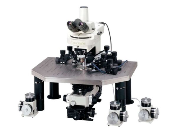
Nikon Instruments Eclipse FN1
The FN1 supports precision imaging with minimal vibration, optimized for electrophysiology studies.
-
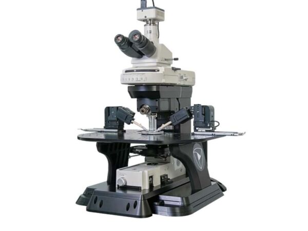
Nikon Instruments Sensapex FN-M
The FN-M combines Nikon optical systems with Sensapex integration for neural imaging precision.