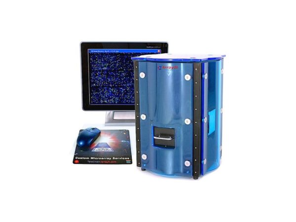
Advanced Microarray Scanners and Accessories: A System Overview for Instruments
Microarray scanners are sophisticated optical instruments engineered for the high-precision imaging and quantitative analysis of microarray slides and chips. The selection of an appropriate scanning system hinges upon critical performance metrics including analytical sensitivity, spatial resolution, scan area, acquisition speed, and overall ease-of-use. A typical instrument is equipped with multiple excitation lasers and a suite of emission filters to accommodate a broad spectrum of fluorescent dyes.read more
The core imaging technology often incorporates confocal optics and dynamic autofocus systems to maintain an optimal focal plane across the entire array, thereby ensuring superior image quality and enhancing the detection of low-abundance molecular targets. For any modern lab, finding a comprehensive solution that includes an automated scanning option is critical for multiplexed assays. Acquired data is then rendered on a digital display for subsequent analysis. Features such as programmable laser-saver functions are also vital for extending instrument lifespan and minimizing maintenance in high-throughput environments.
What Are the Key Specifications and Technology Options for an Advanced Microarray Reader?
- Contemporary microarray scanner systems afford exceptional image clarity, with spatial resolutions as fine as 0.5 micrometers per pixel, facilitating the detailed visualization of high-density microarrays. The integration of confocal optics and dynamic autofocus systems enhances analytical sensitivity and ensures the accurate quantification of low-abundance targets.
- These systems are engineered to support single-, dual-, and triple-color fluorescence scanning, accommodating a wide range of fluorescent labels. This multiplexing capability permits the simultaneous detection of multiple molecular markers, thereby maximizing the data output from a single scan.
- The instrumentation is designed with the versatility to image diverse array types, including DNA, RNA, protein, peptide, glycan, and tissue arrays, on various substrates such as glass, nitrocellulose, or cell-coated slides. This adaptability renders them suitable for a broad spectrum of genomics, proteomics, and biomedical research applications.
- Advanced scanners featuring near-infrared (NIR) fluorescence detection capabilities effectively minimize background signal and autofluorescence originating from substrates like nitrocellulose membranes. This feature significantly improves the signal-to-noise ratio and analytical accuracy, making such systems ideal for quantitative protein and reverse-phase protein array analyses.
- High-throughput functionality is achieved via integrated autoloader accessories, which enable the automated loading and processing of up to 24 slides in a single batch. Automated scanning, image acquisition, and preliminary analysis reduce the need for user intervention and substantially increase productivity for large-scale studies.
- Dedicated analysis software facilitates seamless image acquisition, batch scanning, and data processing with an adjustable dynamic range from 16-bit to 20-bit. Integrated features such as automatic gridding, barcode tracking, and performance validation tools ensure reproducible and traceable results.
- These scanners are constructed for long-term operational stability and minimal maintenance, incorporating features like programmable laser-saving functions to extend laser diode lifespan. Their robust engineering, integrated validation tools, and consistent performance guarantee reliability in demanding research environments.
- Specialized microarray slide scanners, particularly those optimized for DNA arrays, deliver high-resolution and high-sensitivity fluorescence detection. This is essential for precise imaging and quantification, enabling accurate gene expression and genotyping analysis from a single slide.
How is Microarray Analysis Applied Across Key Research Areas?
- Gene Expression Analysis: These instruments are widely employed to profile genome-wide expression patterns by quantitatively comparing messenger RNA (mRNA) levels between physiological states, such as healthy versus diseased tissues. This research elucidates mechanisms of gene regulation, disease pathogenesis, and biological responses to therapeutic interventions.
- Genotyping and Mutation Detection: The technology enables the high-throughput identification of genetic variations, including single-nucleotide polymorphisms (SNPs) and pathogenic mutations associated with both inherited and somatic diseases. High-resolution imaging ensures the precise detection of genetic differences across large sample cohorts.
- Cancer Research and Biomarker Discovery: Microarray scanners are indispensable tools for profiling tumor-specific biomarkers and characterizing aberrant gene expression signatures in cancer cells. These analyses support early diagnosis, the selection of personalized therapies, and fundamental studies of cancer progression and drug resistance mechanisms.
- Pharmacogenomics and Drug Development: In the field of pharmacogenomics, these systems are used to investigate how genetic polymorphisms influence an individual’s response to pharmaceuticals. The resulting data informs the development of safer and more efficacious therapies tailored to a patient’s unique genetic profile.
- Infectious Disease Detection: Scanners are applied to the multiplexed identification of bacteria, viruses, and other pathogenic microorganisms in clinical and environmental samples. This approach allows for the simultaneous detection of numerous infectious agents, thereby improving diagnostic speed and accuracy.
- Toxicology and Environmental Studies: In the discipline of toxicogenomic, these instruments are used to assess the impact of environmental toxins and chemical agents on global gene expression patterns in cells or tissues. Such applications help identify potential public health risks and support regulatory safety assessments.
- Protein, Peptide, and Glycan Analysis: The same scanner platforms are utilized in proteomics and glycobiology to detect and quantify proteins, peptides, and glycans immobilized on microarray slides. These analyses are invaluable for investigating protein-protein interactions, vaccine development, and host-pathogen dynamics.
- Clinical Diagnostics and Personalized Medicine: In clinical settings, microarray scanners play a pivotal role in the diagnosis of genetic and metabolic disorders through the high-throughput screening of patient samples. The data generated directly supports the paradigm of personalized medicine by guiding therapeutic decisions based on an individual’s molecular profile.
Showing 1–12 of 34 results
Category
- Bioreactors+
- Parallel Bioreactors
- Single-Use Bioreactors
- Stirred-Tank Bioreactors
- Capillary Electrophoresis Instruments+
- Capillary Gel Electrophoresis (CGE)
- Capillary Isoelectric Focusing (CIEF)
- Capillary Zone Electrophoresis (CZE)
- Cell Counters and Analyzers+
- Automated Cell Counters
- Manual Cell Counters
- Cellular Imaging Systems+
- Fluorescence Imaging Systems
- High-Content Imaging Systems
- Live-Cell Imaging Systems
- Centrifuges+
- Benchtop Centrifuges
- Clinical Centrifuges
- Micro Centrifuges
- Refrigerated Centrifuges
- Ultracentrifuges
- Chromatography Instruments+
- Gas Chromatography
- Ion Chromatography
- Liquid Chromatography
- Flow Cytometer+
- Cell Analyzers
- Cell Isolation Systems
- Cell Sorters
- High-Throughput Flow Cytometers
- Imaging Flow Cytometry
- Personal Flow Cytometers
- Portable/Benchtop Flow Cytometers
- Spectral Flow Cytometers
- Laboratory Incubators+
- CO2 Incubators
- Dry Bath Incubators
- Hypoxia Incubators and Chambers
- Incubator Shakers
- Laboratory Ovens
- Portable Incubators
- Thermal Mixers
- Laboratory Microscopes+
- Atomic Force Microscopes (AFM)
- Automated Modular Microscopes
- Compound Microscopes
- Confocal Microscopes
- Digital Microscopes
- Fluorescence Microscopes
- Inverted Microscopes
- Live Cell Imaging Microscopes
- Metallurgical Microscopes
- Multiphoton Microscopes
- Polarizing Microscopes
- Raman Microscopes
- Scanning Electron Microscopes (SEM)
- Stereo Microscopes
- Super-Resolution Microscopes
- Transmission Electron Microscopes (TEM)
- Upright Microscopes (Neuroscience)
- Liquid Handlers+
- Automated Pipetting Systems
- Multi-Channel Liquid Handlers
- Robotic Liquid Handlers
- Variable Volume Pipetting Systems
- Mass Spectrometers+
- Ambient Ionization
- APCI MS
- ESI MS
- FT ICR MS
- ICP MS
- Orbi Trap MS
- Quadrupole MS
- Time-of-Flight MS
- Microarray Scanners+
- DNA Microarray Scanners
- Laser-Based Microarray Scanners
- Microplate Readers+
- Absorbance Microplate Readers
- Fluorescence Microplate Readers
- Luminescence Microplate Readers
- Multimode Microplate Readers
- Particle Counters and Analyzers+
- Airborne Particle Counters
- Laser Diffraction Analyzers
- Liquid Particle Counters
- Nanoparticle Analyzers
- Portable Particle Counters
- Remote Particle Counters
Brand
- ACCU-SCOPE
- Agilent
- Airmodus
- Alphasense
- Analytik Jena
- ARGO-HYTOS
- Arrayit
- Aurora Biomed Inc.
- Azure Biosystems
- Baker
- BD Biosciences
- Beckman Coulter
- Beijing Challen Biotechnology
- Binder
- Bio-Techne
- Biobase
- BioRad
- BMG Labtech
- Bruker
- Celestron
- Cellbox
- Centurion Scientific Ltd
- ChemoMetec
- CleanAir
- Cleatech
- Corning
- Countstar
- CS Instruments
- CUBIC
- Cytek Biosciences
- Cytena
- Cytiva
- Data Technologies
- Descase
- Electrolab Biotech
- Eppendorf
- Euromex
- Evident
- Fermex
- Fluke
- Formulatrix
- Getinge
- Gilson
- GPC Bio
- Haier Biomedical
- Hamilton Company
- Hettich
- Hitachi High-Tech
- Hudson Robotics Inc.
- Hydro
- INFORS HT
- Innopsys
- Jasco
- Jeiotech
- JEOL
- Kanomax
- Keyence
- KNAUER
- LAB-KITS
- Labomed
- Labwit
- Leco
- Leica
- Lighthouse
- Logos Biosystems
- LOSI
- Lumex Instruments
- Luminex
- Malvern Panalytical
- Meiji Techno
- Memmert
- Merck
- Mettler-Toledo International Inc.
- Miltenyi Biotec
- Molecular Devices
- Motic
- MP Filtri
- MSE (Medical and Scientific Equipment)
- NCI
- Nikon Instruments
- NuAire
- ORFLO Technologies
- Oxford Instruments
- PAMAS
- Particle Measuring Systems
- Particles Plus
- PBS Biotech
- PCE instruments
- PerkinElmer
- PharSol
- PHCbi
- Prior Scientific
- Promega
- Qiagen NV
- Revvity
- RION
- Sartorius
- SATAKE
- Sciex
- Sebia
- SETA
- Setra
- Shimadzu
- SIBATA
- Sigma Laborzentrifugen GmbH
- Solaris Biotech
- Solida Biotech
- Sony Biotechnology Inc.
- SPT Labtech Ltd
- Stratedigm Inc.
- Sysmex Corporation
- Tecan
- TES
- Thermo Fisher Scientific
- TSI
- Waters
- Western States Machine Company
- ZEISS
- ZETRON
-
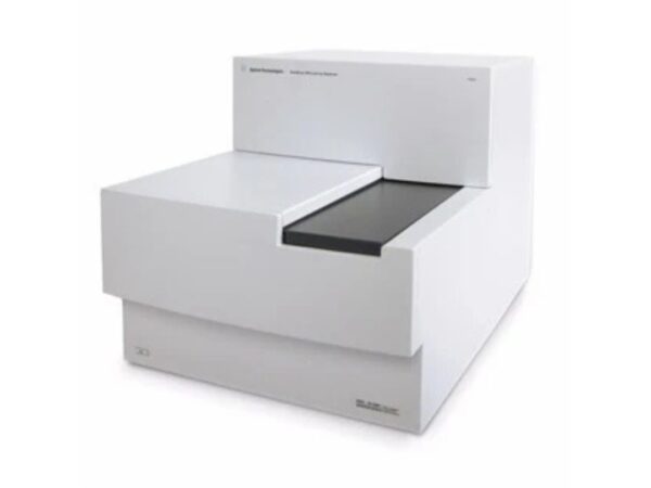
Agilent SureScan
The SureScan Microarray Scanner from Agilent delivers high-resolution scanning for CGH and SNP arrays, providing reliable data for genetic variation s...
-
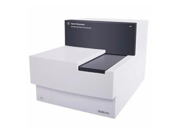
Agilent SureScan Dx
Agilent SureScan Dx Microarray Scanner offers precision scanning for clinical and diagnostic microarray applications, ensuring compliance with IVD sta...
-

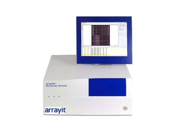
Arrayit Arrayit GenePix
Ensures compatibility with GenePix slides and protocols while providing high-resolution scanning and streamlined integration with fluorescence workflo...
-

Arrayit ArrayPix APXC
Streamlined microplate scanning with fluorescence or absorbance detection, perfect for rapid assay execution in research and pharma labs.
-
Arrayit ArrayPix APXCW
Delivers absorbance, luminescence, and fluorescence reading capabilities for multiple assay formats with intuitive software and fast results across bi...
-

Arrayit ArrayPix APXF
Designed for accurate and efficient scanning of standard microplates, ideal for HTS, drug discovery, and biochemical assay quantification.
-

Arrayit ArrayPix APXFW
Scans multiwell plates (96/384) for fluorescence or absorbance applications, ideal for high-throughput research or pharmaceutical assay screening.
-

Arrayit SpotLight 2 PWSL
Comprehensive support packages covering labor, parts, and diagnostics to ensure continuous uptime and reduce lab delays due to maintenance.
-

Arrayit SpotLight 2 SL2U
Flexible high-resolution scanning system designed to accommodate diverse microarray applications in R&D or diagnostic laboratories.
-

Arrayit SpotLight 2 SLMS
Provides cost-effective, high-resolution scanning of fluorescent microarrays, suitable for education, testing, and low-throughput workflows.
-
Arrayit SpotLight Blue SLMSB
Scanning solution for fluorescence-labeled DNA, RNA, and protein arrays with excellent resolution and sensitivity for research or diagnostic use.
-
Arrayit SpotLight Turbo PWSLT
Enables simultaneous detection of six fluorescent signals, ideal for multiplex experiments, pathway studies, and high-content analysis.